Learn about thermal imaging in veterinary diagnostics, a non-invasive tool for identifying health issues in animals through heat patterns.
Understanding Thermal Imaging in Veterinary Diagnostics
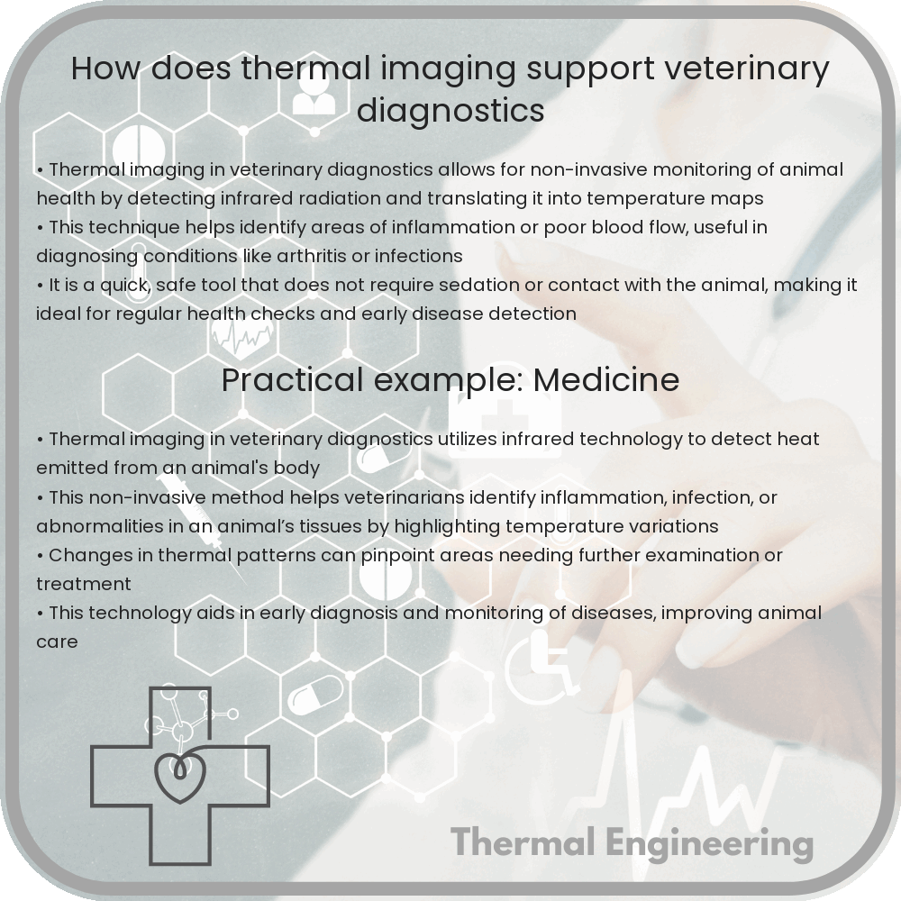
Thermal imaging, also known as thermography, is a powerful diagnostic tool used in veterinary medicine. This non-invasive technique captures and records the infrared radiation emitted from an animal’s body. Essentially, it helps veterinarians visualize the heat distribution across animal tissues, which can be indicative of underlying health issues. Let’s explore how thermal imaging supports veterinary diagnostics and its benefits.
How Thermal Imaging Works
Every object emits infrared energy, which increases with temperature. In veterinary applications, a thermal camera detects this infrared radiation and converts it into an image known as a thermogram. These images display variations in temperature with different colors, typically indicating hotter areas in white or red and cooler areas in blue or purple.
The principle behind this is based on the fact that normal and abnormal tissues emit different amounts of heat. For example, inflamed areas are usually warmer than the surrounding tissues, and areas with poor blood flow are cooler. By interpreting these temperature differences, veterinarians can identify abnormal physiological processes.
Applications of Thermal Imaging in Veterinary Practice
- Detection of Inflammation: Thermal imaging can detect areas of increased heat due to inflammation or infection, which is particularly useful in diagnosing conditions like arthritis, abscesses, or tendonitis.
- Identifying Muscle and Nerve Injuries: Changes in the thermal pattern can indicate muscle or nerve damage. This is critical in sports medicine, helping to diagnose issues in performance animals such as racehorses or working dogs.
- Monitoring Healing: Veterinarians use thermal imaging to monitor the healing process of previously diagnosed injuries or conditions. It provides a clear picture of how the affected area’s heat signature changes over time, which helps in assessing the effectiveness of treatments.
Benefits of Thermal Imaging in Veterinary Diagnostics
Thermal imaging offers significant advantages in veterinary medicine:
- Non-Invasive: It does not require any physical contact, which reduces stress and discomfort for animals during examinations.
- Safe: This method does not involve ionizing radiation, making it safe for repeated use, which is particularly important for monitoring chronic conditions or recovery processes.
- Instant Results: Thermal images are produced in real-time, which allows for immediate assessment and aids in making quick medical decisions.
- Comprehensive: It can evaluate large areas of the body quickly, providing a broad picture of the animal’s physiological state.
Although thermal imaging is a valuable tool in veterinary diagnostics, it should be noted that it is typically used in conjunction with other diagnostic methods, such as X-rays, ultrasound, or blood tests, to confirm findings and obtain a comprehensive view of an animal’s health.
Challenges and Considerations
Despite its many benefits, thermal imaging requires proper understanding and interpretation. The accuracy of the results depends on various factors, such as the ambient temperature, animal’s fur, and the skill of the technician. Incorrect interpretation can lead to misdiagnosis. Therefore, specialized training for veterinarians and technicians in thermography is crucial.
In conclusion, thermal imaging is a transformative technology in veterinary diagnostics, providing quick, safe, and non-invasive evaluations of animal health. As technology advances and becomes more accessible, it holds the potential for even broader applications in veterinary medicine.